Bill’s Vitrectomy
A photo essay by Bill Leahy Jr.
| Bill Leahy Jr. is a lecturer at the College of Computing, Georgia Institute of Technology. He has kindly given us permission to publish his excellent account of a vitrectomy performed on one of his eyes several years ago. Warning: These photos may be disturbing to some people.Rest assured, however, that the procedure is safe and painless.Our thanks to Bill for sharing his experience with us. If you would like to visit his personal home page, where this information also appears, follow this link. For more information about eye anatomy, see Anatomy of the Eye. To read another personal account (no photos) of this procedure, see Retinal Detachment and Vitrectomy Surgery. |
In December 1997 I had eye surgery to correct a condition where some blood vessels in my eye had ruptured. The surgery is basically intended to remove any blood remaining in the clear jelly or vitreous humor which fills the eye. Once the blood is removed a laser is used to cauterize any damaged areas to prevent further bleeding.
What was most interesting about the surgery is that for most of it I was awake! The eye to be operated on is anethesized and does not function. The other eye is covered so nothing can be seen. One can, however talk to the surgeon.
They do administer tranquilizers so it is a relatively calm time. There was no pain and actually no significant discomfort. The hospital stay is technically less than 24 hours.
The indescribeable part of the whole process are the images that appear after the surgery in the eye that has been cauterized. The following photos were extracted from a videotape of the surgery that was made using a camera mounted to the microscope used by the surgeons.
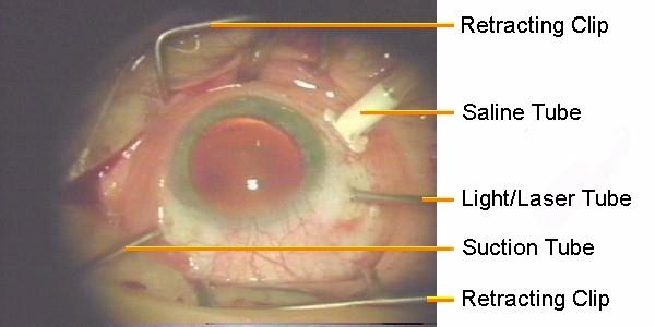
This labeled photo will give an overview of the situation. The eye is held open with retractor clips. A saline line has been stiched to the eyes forcing saline solution into the eyes to maintain the shape and replace any of the vitreous humor that is sucked out with the blood. It also replaces that which simply leaks out of the various incisions in the eye. Two tubes are also inserted through holes in the eye: one is the cauterizing laser which also has a light source for general illumination. The other is the suction tube.

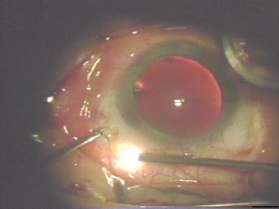
Here one can see clearly the tube penetrating the eye visible though the pupil.
The laser/light tube can be seen here external to the eye in light mode.

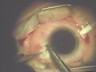
After addition of fluid to lubricate the eye, a lens is placed directly on the eyeball to allow better visibiity of the interior.

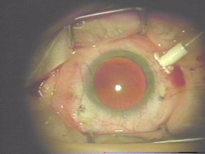
During actual operations on the interior the room lights are dimmed to allow a better view of the interior. All light comes from the light tube. In this photo the blood is actually being sucked out although it is difficult to see in this still photo.

Here the bright spot of light is the actual laser beam during cauterization.

Once the procedure is complete the holes which allowed penetration of the various tubes are temprarily sealed with rivet like metal pieces which will allow the eye to stabilize before suturing.
Here the left access hole is being closed with tiny sutures.
Here both penetration holes have been sutured shut although the saline tube is still in place.

The sutures holding the saline tube have been cut and if you look closely you can see the jet of saline solution still coming out of the tube.